Achilles Tendon Injury
Achilles Tendon Scanning
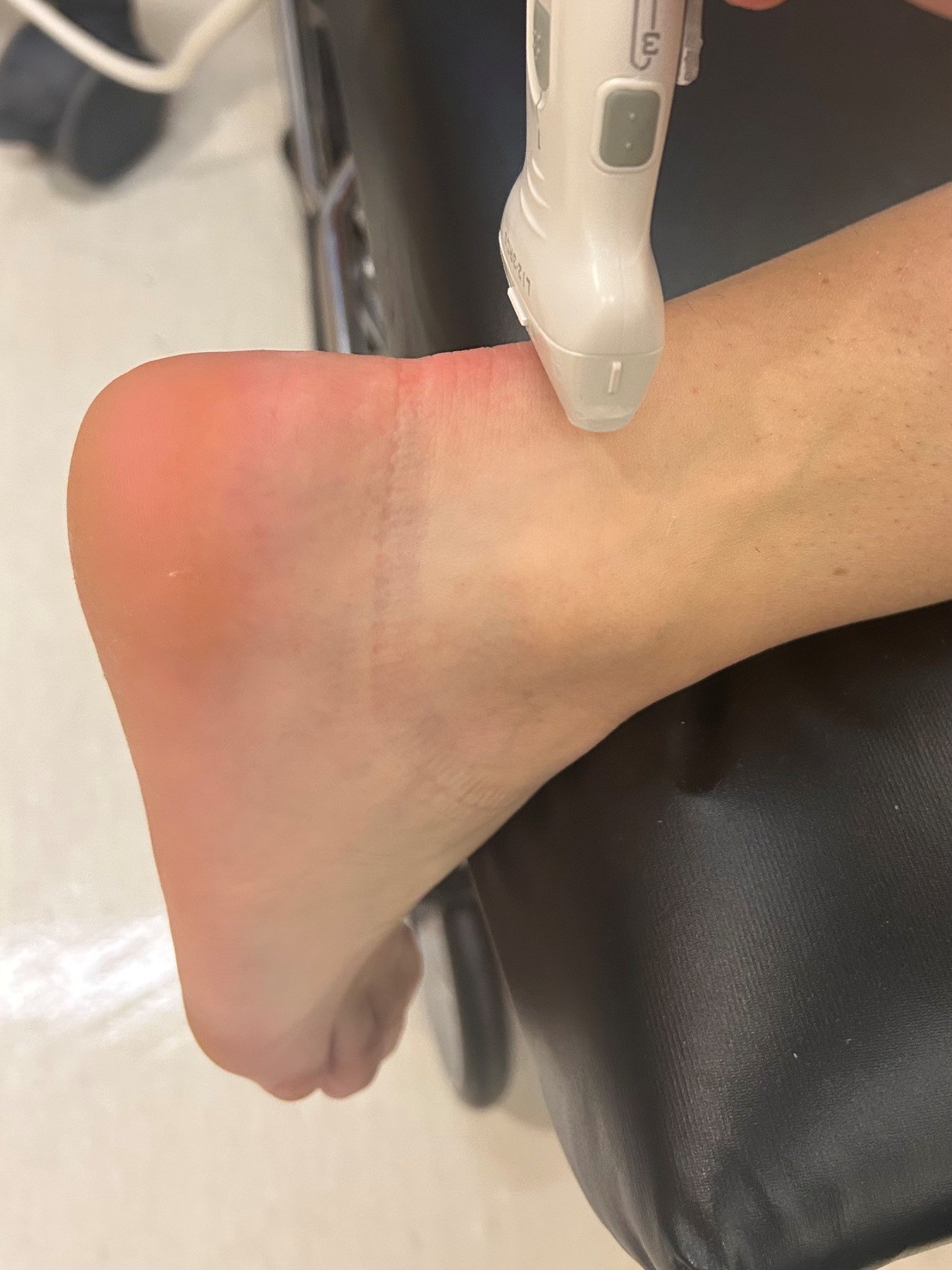
How to scan:
Have the patient lay prone with the foot facing downwards off the side of the bed. Use the linear transducer in sagittal orientation with the probe marker facing cranially. Scan from the calcaneus to visualize insertion, to the body of the gastrocnemius. Switch to transverse orientation with the probe marker facing the patient’s right and repeat. Scan from insertion at the calcaneus to the body of the gastrocnemius.
A meta-analysis of 15 studies (808 patients) found ultrasound has a sensitivity of 94.8% (95% CI 91.3–97.2%) and specificity of 98.7% (95% CI 97.0–99.6%) for detecting complete Achilles tendon ruptures, using surgical confirmation as the reference standard. (1)
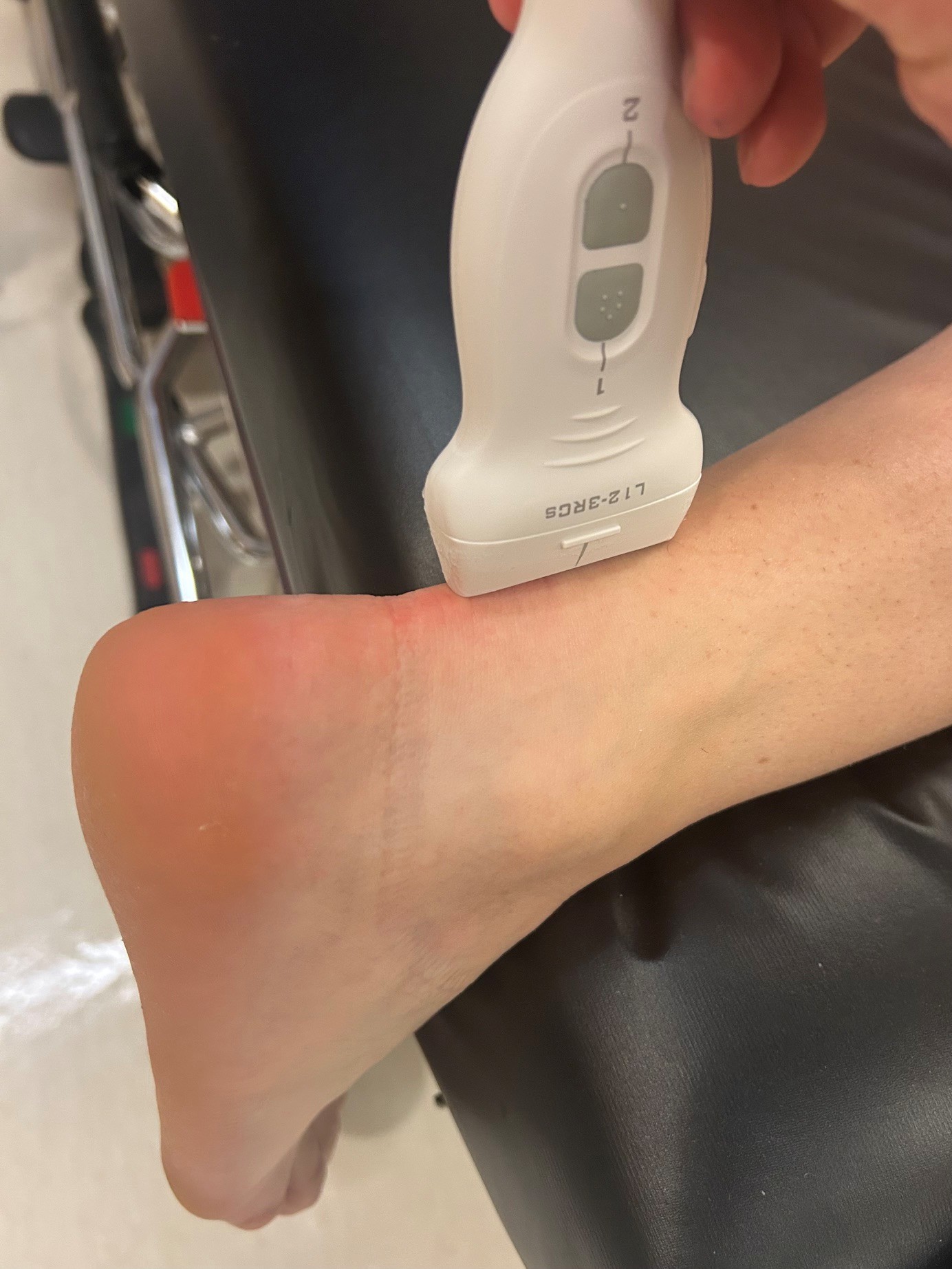
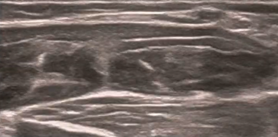
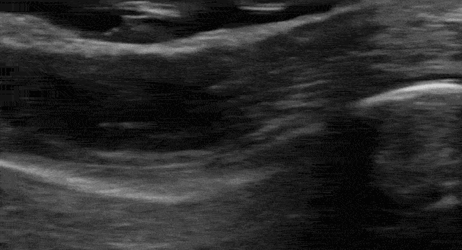
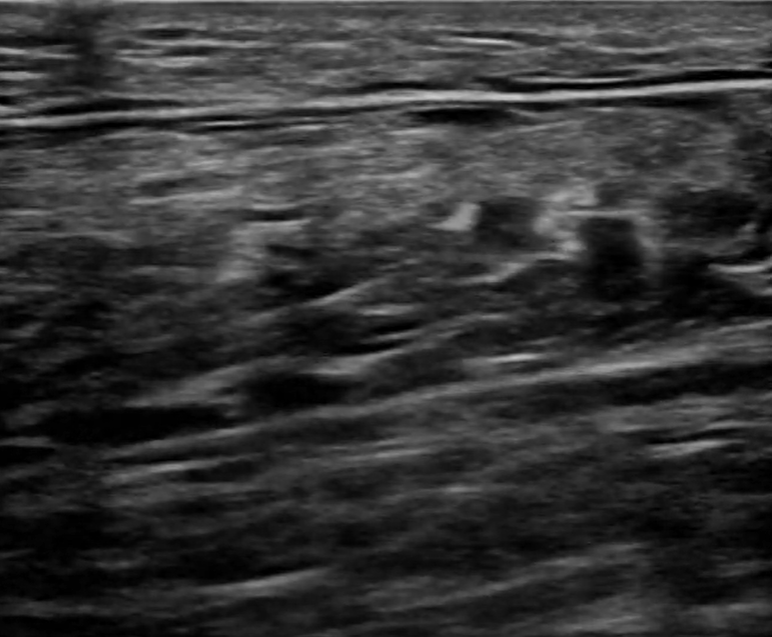
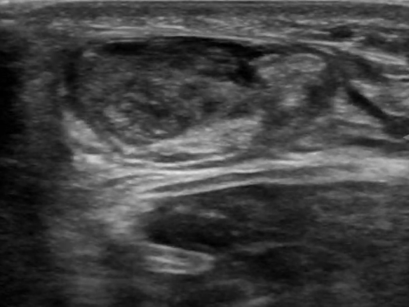
Sagittal scanning
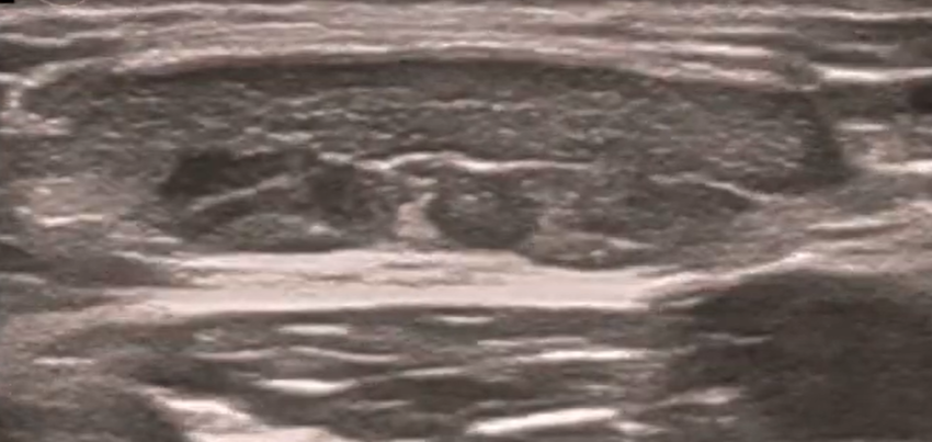
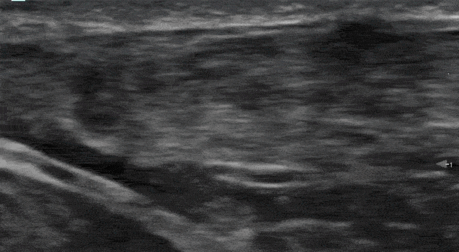
Transverse Scanning
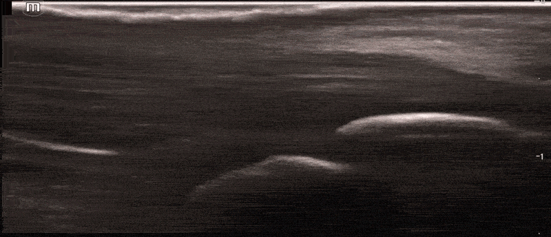
![]()
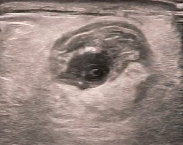
Try to identify the structures demonstrated in the below images.
Click here to see labeled images.
Special tests:
Thompson test-
Flex the knee to 90° and squeeze the patient’s calf. An intact Achilles tendon will fully plantarflex the foot when the calf is squeezed. This is a negative Thompson test.
Pathologic Achilles
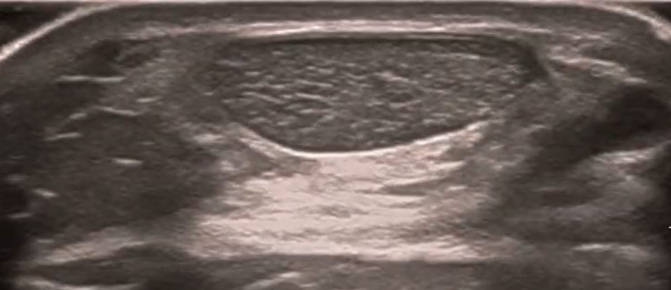
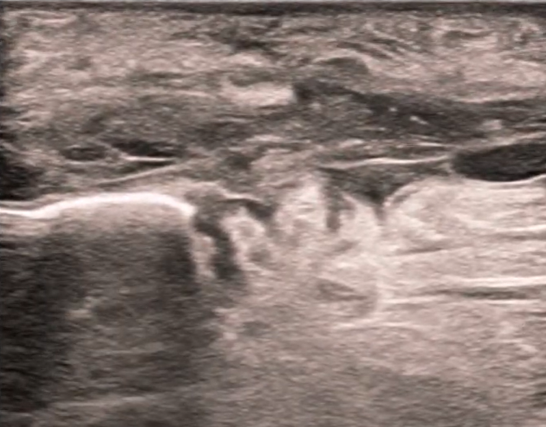
Sagittal scanning
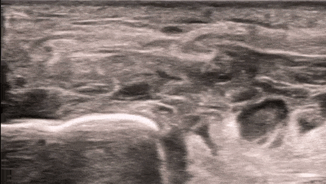
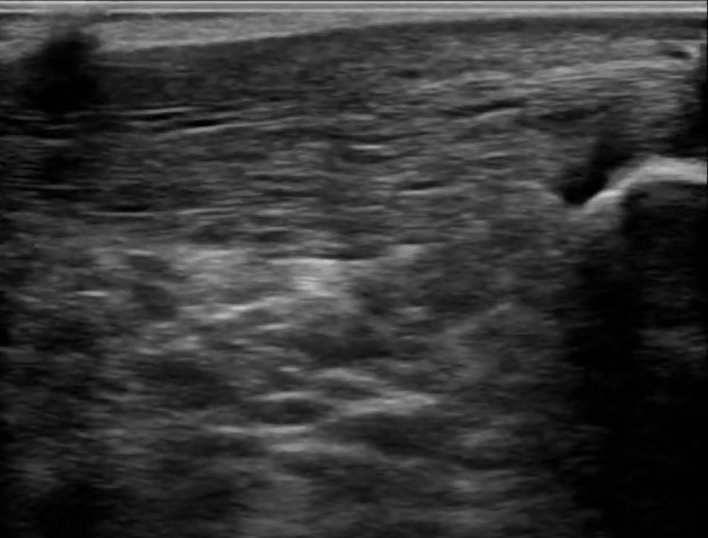
Transverse Scanning
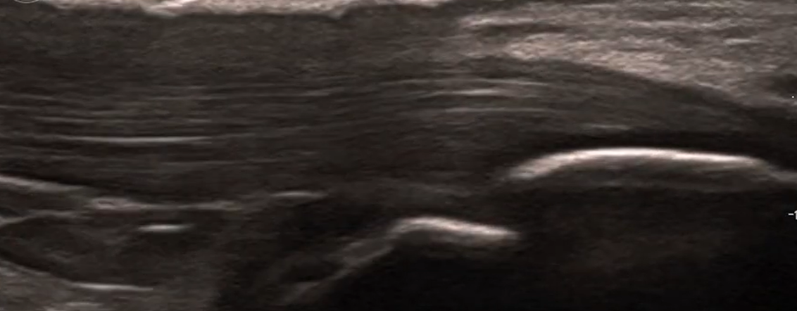
![]()
![]()
Try to identify the structures demonstrated in the below images.
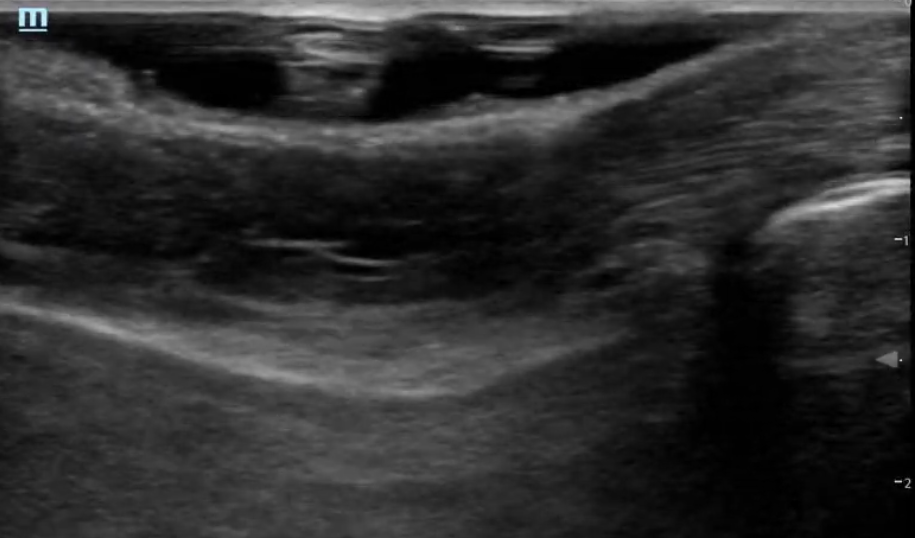
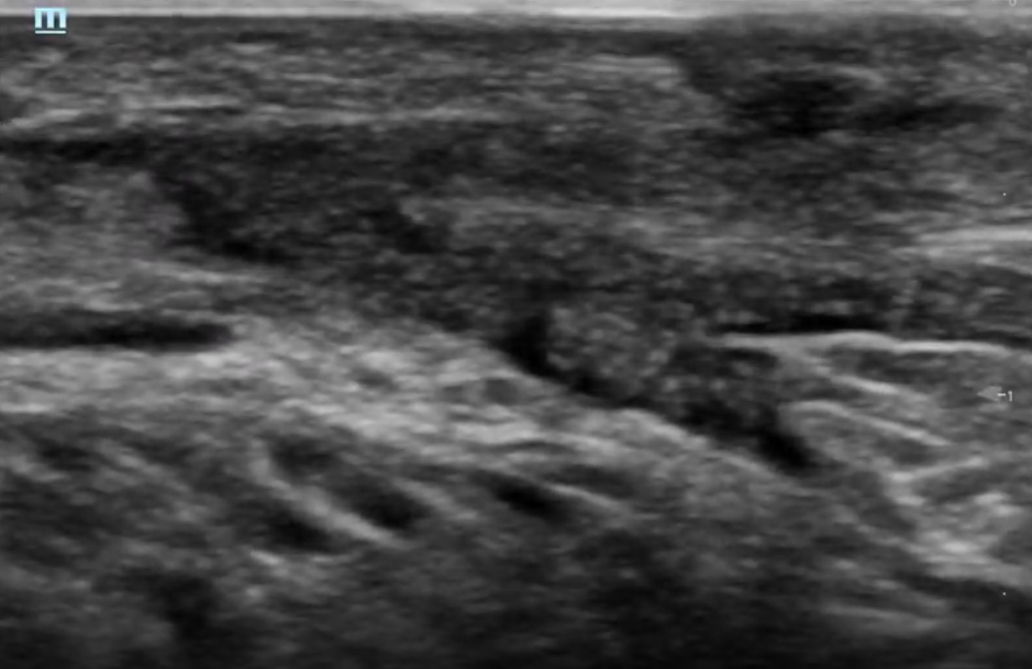
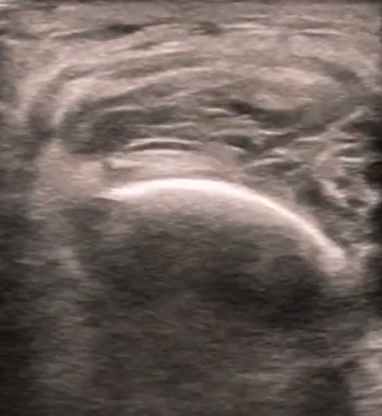
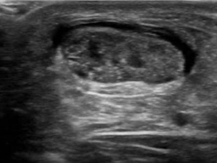
Click here to see labeled images.
Special tests:
Thompson test-
Flex the knee to 90° while lying prone and squeeze the patient’s lower leg. The Thompson test (calf squeeze test) has a sensitivity of 96% and specificity of 93% for complete Achilles tendon rupture, using surgical findings as the reference standard. A positive test is defined as loss of plantar flexion when the calf is squeezed with the patient prone. (2,3)
Visit our case file for more information about the presentation and management of Achilles rupture!
Written by: Meghan Dillan, MD and Thomas Scott, MD
Special thanks to Dr. David Haidar for the provided ultrasound images.
References:
1. Aminlari A, Stone J, McKee R, et al. Diagnosing Achilles Tendon Rupture With Ultrasound in Patients Treated Surgically: A Systematic Review and Meta-Analysis. J Emerg Med. 2021;61(5):558-567. PMID: 34801318.
2. Maffulli, Nicola. The clinical diagnosis of subcutaneous tear of the Achilles tendon. The American journal of sports medicine 26.2 (1998): 266-270.
3. Reiman M, Burgi C, Strube E, et al. The Utility of Clinical Measures for the Diagnosis of Achilles Tendon Injuries: A Systematic Review With Meta-Analysis. J Athl Train. 2014;49(6):820-829. PMID: 25243736.